MANILA – “Kind of spooky” is how Siemens Healthineers Asia-Pacific President Elisabeth Staudinger describes cinematic rendering, the latest technology they are offering to make CT scan images more realistic than ever.
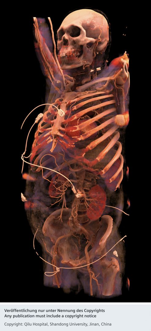
On Wednesday, at a roundtable discussion in Shangri-La at the Fort, Staudinger presented images generated by the newly developed Cinematic Volume Rendering Technique.
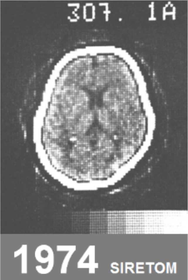
In contrast to a blurry, pixellated, black and white image of the brain from a CT scan in 1974, the video Staudinger showed was filled with colored, detailed parts of the body worthy of a medical drama or the opening credits of a Netflix superhero show.

This technology was actually built by Siemens Healthineers – the healthcare business of Siemens AG – after Hollywood representatives approached them, looking for medical images that were more life-like.
Siemens Healthineers then collaborated with people from the film industry to create the technology, which used images coming from CT scans.
Layer by layer of the body could be stripped away through cinematic rendering. Muscles, bones, organs, vessels, and the like could be viewed in high definition. Users could zoom in and out, and rotate the images, too.
“Looking at the vessels in the brain, imagine a stroke where you would want to know, is it a clogged vessel or did the vessel burst? It’s something you would easily be able to see,” Staudinger said. “Kind of spooky in a way, right?”
Cinematic rendering was also now being used in medical schools, so students could learn about the human anatomy without needing a cadaver, said Siemens Healthineers Philippines President Mike Tan.

The technology made the parts of the body more visible. However, Staudinger acknowledged, it did not yet tell the user what the patient’s problem was.
Next question: so, what’s patient’s problem?
“And this is now the next step, is what you can put on top of the already existing image information, and then you can train with artificial intelligence algorithms. You can train the computer, more or less, to look for problems in an image,” she said.
How does one train a computer, then?
“You feed many, many images,” Staudinger explained. “So you do, for instance, a lung scan, and then you have hundreds or thousands or tens of thousands of lung scans in a database. And in the beginning, of course, you need to guide the computer. You need radiologists to annotate those images and say, ‘Look, this is abnormal,’ ‘This is a tumor,’ or, ‘If you find something like this, it is suspicious of that.’ And then you feed the computer with images and they use this knowledge you gave them in the beginning and they try to refine it and then the computer itself will be able to show you, ‘Well, maybe you can check that section in the body, this does not look normal.'”
This is what Siemens Healthineers is now developing together with researchers from universities, hospitals, and other partners worldwide.
“Because you need to… train the computer to know what the machine is looking at. And in the end, this will benefit the patient in the sense that it will be faster to diagnose, and the diagnosis will be more accurate. There’s just less error,” she explained.
Staudinger added, “If you combine the knowledge of the radiologist with the support of such algorithms, you can really improve the accuracy and the precision of the diagnosis.”
According to a press release, cinematic rendering works by “calculating realistic lighting effects.”
“For instance, the depth of a fracture is fed into the calculations. The deeper the fracture, the less light is able to penetrate, resulting in a range of shadows. The result is an almost perfect depiction of fractures, ivory-colored bones, and clearly defined organs and blood vessels – easily discernible from each other thanks to the inclusion of shadows and depth,” the press release says.


